Drawing Blood – the Origins Story
‘Drawing Blood’ emerged from a teaching session I inherited on ‘Blood abnormalities’. Let’s be clear at the outset, I’m not trained as a haematologist or even as a hematologist. Why did this end up with me? Well this is the beauty of the fundamentals of cell biology: knowing these gives you considerable insight into how any cell functions. It’s not my job to teach the clinical application, that’s what the medical doctors do. Conversely, with all due respect, few practising clinicians are going to be able to explain the finer details of cytoskeletal function. I was already well aware of certain parallels, for example immune cells can form a synapse using many of the same proteins I studied in neuronal synapses. The further I dived into this new topic the more intriguing examples I found. The formation of platelets relies on a process of microtubule extrusion very similar to one I’d investigated in growing neuronal axons that I wish I’d been aware of at the time. Good old hindsight. The second thing that grabbed my attention was the diversity of red blood cell shapes and the varied, descriptive names given to them. This set me off down internet rabbit holes finding as many ‘-cytes’ as I could. You can see a spotter’s guide to them on the erythrocyte page.
Seeing red…circles
At first glance, the small, regular discs of red blood cells were not the most obvious inspiration for a series of cartoons. Certainly not when compared to the sweeping diversity in size and shape of brain cells. Reading more about red cell variations, hinted at ways to emphasise their distinctions in cartoon forms while still retaining an image that communicated factual understanding of their biomedical relevance. Indeed, one of the foundations of cartooning is being able to capture the essence of an individual or object with an economy of detail. Everyone knows Snoopy is a dog but he’s probably not the best way to learn canine anatomy if you’re a budding vet. Similarly, a good example of diverse personalities based on a simple body plan are the Minions.


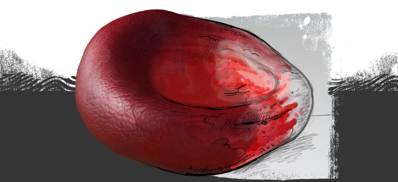
One of the important physiological features of red blood cells is their three dimensional shape. Normal ones are a biconcave disc – think of orecchiette pasta or a ball of clay squashed between thumb and forefinger – 7 microns in diameter, with such consistency they can be used as a tiny ruler when looking down a microscope. This shape allows the optimal surface area for gas exchange so they can carry and release oxygen efficiently around the body. At the same time they have to be deformable enough to squeeze through the tiniest capillary (sometimes less than 7 microns wide!) and tough enough to withstand being blasted through arteries. Many of the shape variations illustrated here are lined to pathologies precisely because the change in shape impairs their function in some way. Underlying this, these shape changes have many causes: genes, metabolism, diet, toxins, environment. Understanding the links from this molecular level, to the shape changes, to effects on oxygen delivery and thus health is clearly crucial. Typically though, this 3D element is lost. Textbooks rarely show it and in clinical practice blood smears are an invaluable tool but as the name suggests the cells are flattened across a slide and viewed in a 2D microscope image. This set me off on another journey of artistic and technological discovery in the world of 3D computer graphics. I had already started this in the context of teaching protein function (I’ve made a start on my story of 3D models) and this suggested a way to use it at a cellular rather than molecular scale. After a lot of false starts negotiating what felt a daunting learning curve, I made 3D versions of the entire spotter’s guide. You can see these at my Sketchfab site. Shortly afterwards, I spotted a notice in the Graphic Medicine newsletter asking for contributions to something called The Blood Project. Sometimes Fate smacks you in the face with a great big fish and you need to take the hint. I was thrilled when they liked my blood images and used them in their Visual Art resources on schistocytes and granulocytes. Even better, I was a guest on their podcast hosted by Helen Osborne, you can listen here to ‘Follow your Curiosity: Humanities in Science‘ or search on Spotify for ‘The Blood Project’. This was great fun talking about how I’ve used art throughout my research, outreach and teaching and will hopefully inspire others to do likewise. It’ll take me ages to fill in all the scientific details for each of the cells on the erythrocyte page so if you want to find out more The Blood Project is a great place to start.
Mixed reality
I’m pleased with my 3D models of blood cells, although the more I learn about the digital techniques, the more I think I should go back and refine them… Anyhow, it struck me that in an ideal world someone would be able to see the 3D rendering alongside the cartoon or a 2D clinical image. This led to, yes, another rabbit hole with a steep learning curve and mixed metaphors, this time of Augmented Reality (AR). Many people are familiar with Virtual Reality (VR) however this relies on expensive headsets which isolate the viewer from the real world. AR recreates the sense of 3D immersion by overlaying virtual objects on physical ones so the experience can be shared. I’ll write some more later about how you can use apps to recreate this for yourself for now here are some films of how I’ve begun to use it. One shows the Red Cell Spotters Guide, if you hold a phone, iPad or tablet over the poster, the software recognises the cartoon cell and projects the relevant 3D virtual model over it. The second example uses the Wordotomy card game I co-created. I’m developing an AR version of the entire deck but for now here is the ‘eryth-‘ card brought to life. See how cool it is when the cells rotate with the card!
After the Red Army
There are many more cells found in the blood. The next subject, at least as far as my teaching was concerned, was platelets. ‘White’ blood cells refers to a range of immune cells and it was difficult to know where to begin. They’re not even white and many of their traditional names are historical artefacts of the early chemical stains used to visualise them and bear limited connection to their actual function. Modern molecular techniques are constantly revealing new intricacies and diversity in the roles and identities of these cells which can change over their lifespan. I had already done some drawings of macrophages, large cells which digest invading pathogens or defunct host cells, so this seemed a good place to begin. Their behaviours readily lent themselves to cartoon ideas which helped. Next up were the granulocytes, a relatively well-defined group which, with their involvement in allergies and fighting different bugs, were promising material.
And this is where I’m up to. I’ve not started on B cells making antibodies, never mind T cells with all their subdivisions of cytotoxic and helper cells… Please visit the pages for platelets, macrophages and granulocytes to find more cartoons and information about the cells featured: